

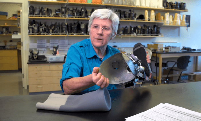
Researchers at the University of Washington have developed a more efficient method of manufacturing transtibial prosthetic sockets with embedded sensors capable of tracking movement between the residual limb and socket.
The work, highlighted in a recent EurekAlert news release, addresses one of the most persistent problems in lower-limb prosthetics: socket fit changes over time, but clinicians have limited objective information about what happens inside the socket once a patient leaves the clinic.
Led by Professor Joan E. Sanders, the research team created a hybrid manufacturing process that combines a 3D-printed inner structure, embedded sensing components and an external carbon-fibre lamination. The resulting socket can continuously monitor relative limb motion while protecting the electronics beneath the socket wall.
The findings were published in the peer-reviewed journal Frontiers in Rehabilitation Sciences.
Moving socket-fit assessment beyond the consultation room
Socket fit is not static. Residual-limb volume, tissue position, activity, suspension and physiological changes can alter the relationship between a person’s limb and prosthesis throughout the day.
Patients may report looseness, pressure, instability or discomfort, but these descriptions do not always reveal precisely when or where a change is occurring. A socket that appears satisfactory during a short clinical appointment may behave differently during prolonged walking, work, travel or other daily activities.
In a Frontiers interview about her research, Sanders said her team is attempting to replace some of this uncertainty with continuously collected quantitative information.
The objective is not to replace patient feedback or clinical judgement. Instead, embedded sensors could give prosthetists an additional source of evidence showing how the limb moves within the socket during everyday use.
How the sensor-integrated socket is manufactured
The method described in the original research paper begins with a digital model of the patient’s existing socket. Where a digital file is unavailable, the socket can be scanned to capture its internal shape.
Researchers then create a 3D model of a two-millimetre-thick inner structure. Debossed channels and enclosures are incorporated into its external surface to accommodate sensor antennae, wiring, electronics and the electrical connector.
The process has three principal stages:
- Producing the customised 3D-printed inner structure
- Installing the sensing harness within the prepared channels
- Applying a carbon-fibre lamination for structural reinforcement
The sensors are positioned between the printed structure and outer laminate. Unlike pressure sensors placed directly against the residual limb or liner, the system measures relative motion without requiring direct contact with the skin-facing surface.
This protects the electronic components from mechanical damage and allows measurements during both stance and swing phases of walking.
Fabrication time reduced by 12%
The researchers reported that an experienced technician required 730 minutes to manufacture the new sensor-integrated socket, compared with 830 minutes for the team’s traditional method of producing a socket with sensors.
This represents a 12% reduction in total fabrication and curing time. The relevant comparison is between two sensor-equipped socket processes—not between the investigational socket and a standard clinical socket without sensors.
The team believes further automation could eventually produce larger time savings. Automating the placement of sensor channels and component housings within the CAD model, for example, could substantially reduce digital preparation time. Prefabricated sensor harnesses could also replace components that currently need to be assembled individually.
These additional savings remain projections and have not yet been clinically validated.
Early testing identifies both potential and risk
The study included four transtibial prosthesis users aged between 42 and 65. All had experienced traumatic amputation and were classified as at least K2 ambulators. Participants used the investigational sockets in their home and community environments for periods of up to four weeks.
The sockets increased in volume by less than 0.2% during take-home use, suggesting relatively limited deformation over the testing period. Mechanical samples were also subjected to 300,000 loading cycles without meaningful change in shape.
However, the small study also demonstrated why manufacturing accuracy and individual clinical review remain essential.
Two participants noticed that the normal 3D-printed surface was rougher than their conventional socket. One felt that the additional friction improved suspension and reduced pistoning; another found that the socket adhered too strongly to the liner and felt heavier.
One participant developed a patellar blister after the printed inner structure failed to reproduce important socket contours accurately. The user stopped wearing the investigational prosthesis, and the research prosthetist followed the participant during recovery.
The researchers subsequently tested a vapour-smoothed internal surface. This produced friction characteristics closer to those of a conventional laminated socket and was viewed more favourably by one participant. The findings indicate that surface finish cannot be treated merely as a cosmetic manufacturing detail: it can directly affect donning, suspension, perceived weight, movement and skin health.
Monitoring is not yet automatic adjustment
The present study demonstrates a method for manufacturing sockets with protected embedded sensors. It does not establish a commercially available socket that automatically changes shape in response to the collected data.
Sanders and her colleagues have previously investigated adjustable sockets and feedback-controlled systems. Their longer-term vision is for sensor information to help clinicians recognise deteriorating fit and, eventually, support sockets that adapt to residual-limb changes in real time.
Before this becomes routine clinical practice, researchers must determine which measurements are genuinely useful. Detecting pistoning or changes in anterior-distal motion is technically possible, but clinicians need validated thresholds that indicate when an adjustment, sock change, suspension review or socket replacement is appropriate.
More data do not automatically produce better care. Sensor readings must be reliable, interpretable and connected to decisions that improve comfort, limb health, mobility and participation.
Potential relevance across the IMEA region
Sensor-integrated sockets could be particularly relevant to prosthetic services across the Middle East, Africa, Central Asia and South Asia, where patients may travel considerable distances for follow-up and specialist prosthetists may cover large geographical areas.
If validated for longer-term use, remote socket monitoring could help clinical teams identify emerging fit problems between appointments. It could also support more informed follow-up for patients returning to work, living in rural communities or receiving services through outreach and humanitarian programmes.
The hybrid manufacturing model may also be relevant. A specialist facility could produce the printed inner structure and sensor harness, while regional prosthetic centres complete fitting, alignment, lamination and rehabilitation. Such a model could connect digital central fabrication with locally delivered clinical care.
Implementation would still require:
- Accurate scanning and digital socket-design capability
- Reliable printing and quality-control standards
- Trained prosthetists and technicians
- Sensor calibration and maintenance procedures
- Secure systems for storing and reviewing patient data
- Clear responsibilities for remote clinical monitoring
- Access to replacement electronics and repair services
Cost will also be critical. The study examined fabrication efficiency but did not establish the commercial cost of the socket, sensors, software or long-term monitoring service.
Data should strengthen the patient–prosthetist relationship
Sanders’ research presents the smart socket as a clinical tool rather than a substitute for professional care. That distinction is important.
A patient’s account of comfort, confidence and function remains essential, while the prosthetist must interpret the socket within the wider context of residual-limb health, alignment, activity, components and rehabilitation goals. Sensors may help explain what is happening, but they cannot independently decide what matters most to the person using the prosthesis.
The new fabrication method is therefore best viewed as an enabling step. It makes long-term sensor integration more practical and creates a platform for larger clinical studies. Its ultimate value will depend on whether the collected data lead to earlier intervention, fewer skin problems, more stable fit and better participation in everyday life.
- EurekAlert: Joan Sanders and the challenge of prosthetic socket fit
- Frontiers: Interview with Professor Joan Sanders
- Original research: Quick fabrication of 3D-printed prosthetic sockets with embedded sensors
- University of Washington: Professor Joan Sanders
- IMEA CPO: Adjustable-Volume Sockets Linked to Better Comfort, Mobility and Quality of Life
- IMEA CPO: Light-Emitting Textile Sensors Could Give Prosthetists a New View Inside the Socket
- IMEA CPO: Meablex Develops Integrated Bionic Arm with Adjustable Socket and Magnetic Muscle Sensing